Assignment 2/3 Report
1. Introduction and overview
This augmented reality app is an immersive exploration of diverse cell types, including plant, animal, bacterial, and fungal cells. Its main goal is to enhance understanding cellular structures by visually interactive 3D models and compartments. It also enhances understanding by learning similar but different cellular components of plant and animal cells with 3D models and running videos.
2. Description of the application
The goal of this augmented reality application is to understand four complex 3D cell structures and the differences between plant and animal cells. To begin, 3D models of animal, plant, fungal, and bacterial cells can be displayed on screen by students who can touch virtual buttons to navigate to the 3D models, showing the different arrangements of cell components. Other papers that have animal cell plant cell including cell components can be explored with 3D models and videos can be played for more detailed learning. This innovative approach bridges the gap between traditional 2D materials and the dynamic reality of cells, increasing engagement and understanding.
It is significant that we have solved the difficulty of visualizing cellular structures in a conventional way. In the traditional way, students may feel bored when studying cells only in 2D. This augmented reality solution enhances learning and promotes active participation. Through hardware such as webcams, interactive software features, and usability components, the benefits of augmented reality, such as visual augmentation and tactile interaction, achieve the goals of the application by intuitively engaging users.
3. Interactive design
Students can receive an immersive and personalized education through interaction that goes beyond conventional teaching techniques. Students' learning outcomes are enhanced and their grasp of cytology is deepened by actively engaging in cellular structures and functions.
A piece of paper marked with cells is placed in front of the webcam when the user activates the augmented reality. When the user presses the virtual button, rotating 3D models of bacterial, fungal, and plant cells emerge on the screen. The user then activates 3D models of both plant and animal cells side by side on the paper with the additional cell markers. To contrast the two cell types, the user highlights certain structural elements in each. To further their comprehension, the user can directly engage while viewing rotating cell components. By pressing virtual buttons, the user can also watch and hear an in-depth video and explanation.
4. Technical development
This augmented reality application provides students with an immersive opportunity to engage with dynamic 3D models of cellular structures via webcam. This innovative augmented reality application also provides an interactive learning experience that goes beyond the limitations of traditional 2D resources.
Users can easily explore complex cellular structures with a simple tap of a 3D model manipulation that would be difficult to understand in 2D. In addition, two different cell types can be placed side by side for a comprehensive view that not only captures attention but also helps to understand the multifaceted nature of cellular components. This approach recognizes the unique properties of different cellular structures and promotes more insightful comparative analysis.
For those seeking a more comprehensive understanding, the app offers a variety of informational videos and explanations that can be viewed by tapping the More Virtual button to delve into the intricate details of animal and plant cellular components. Flexible engagement pace and seamless rotation and video playback allow students to customize their learning experience to their liking.
5. Descriptions of 3D model
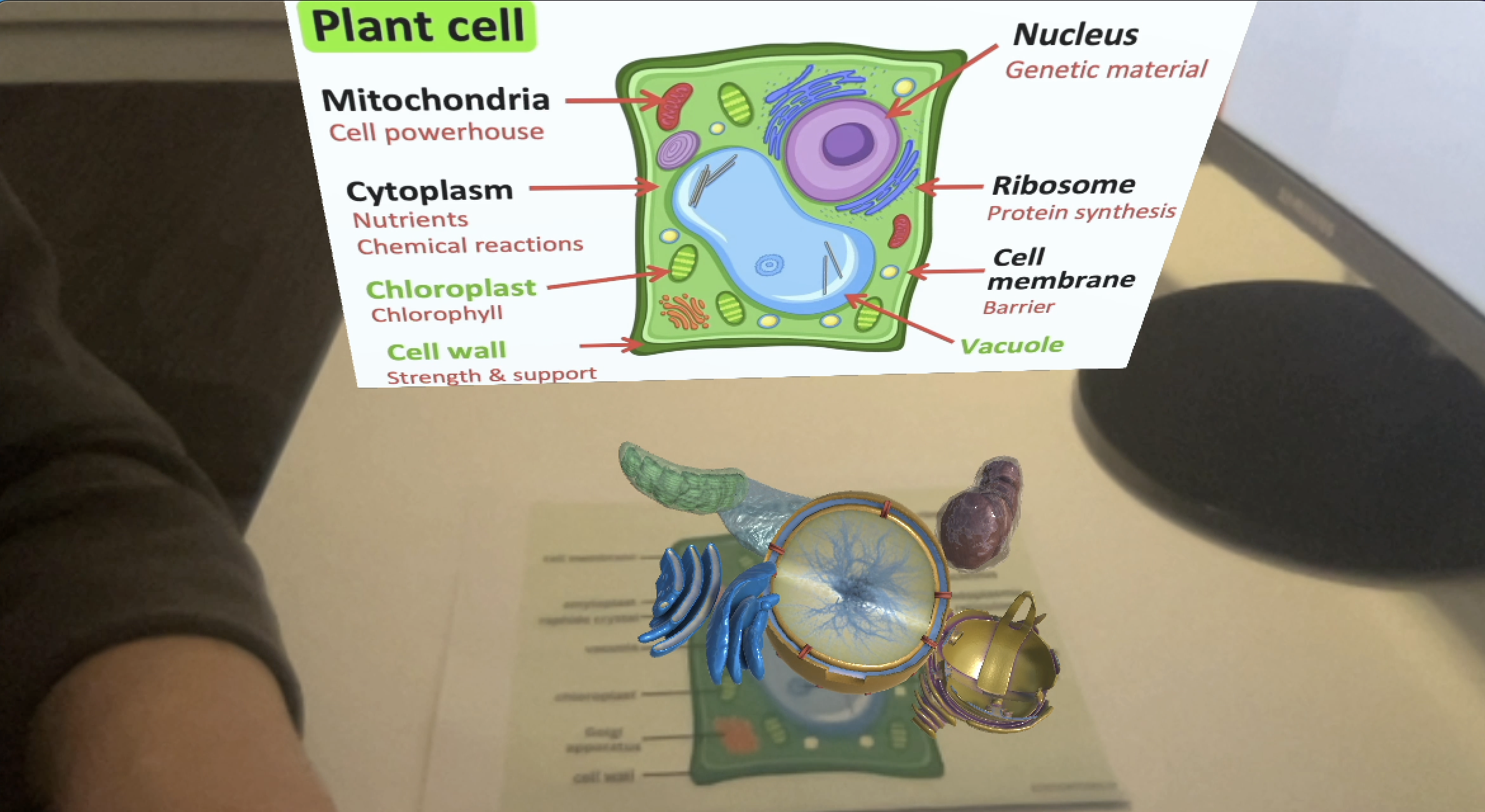
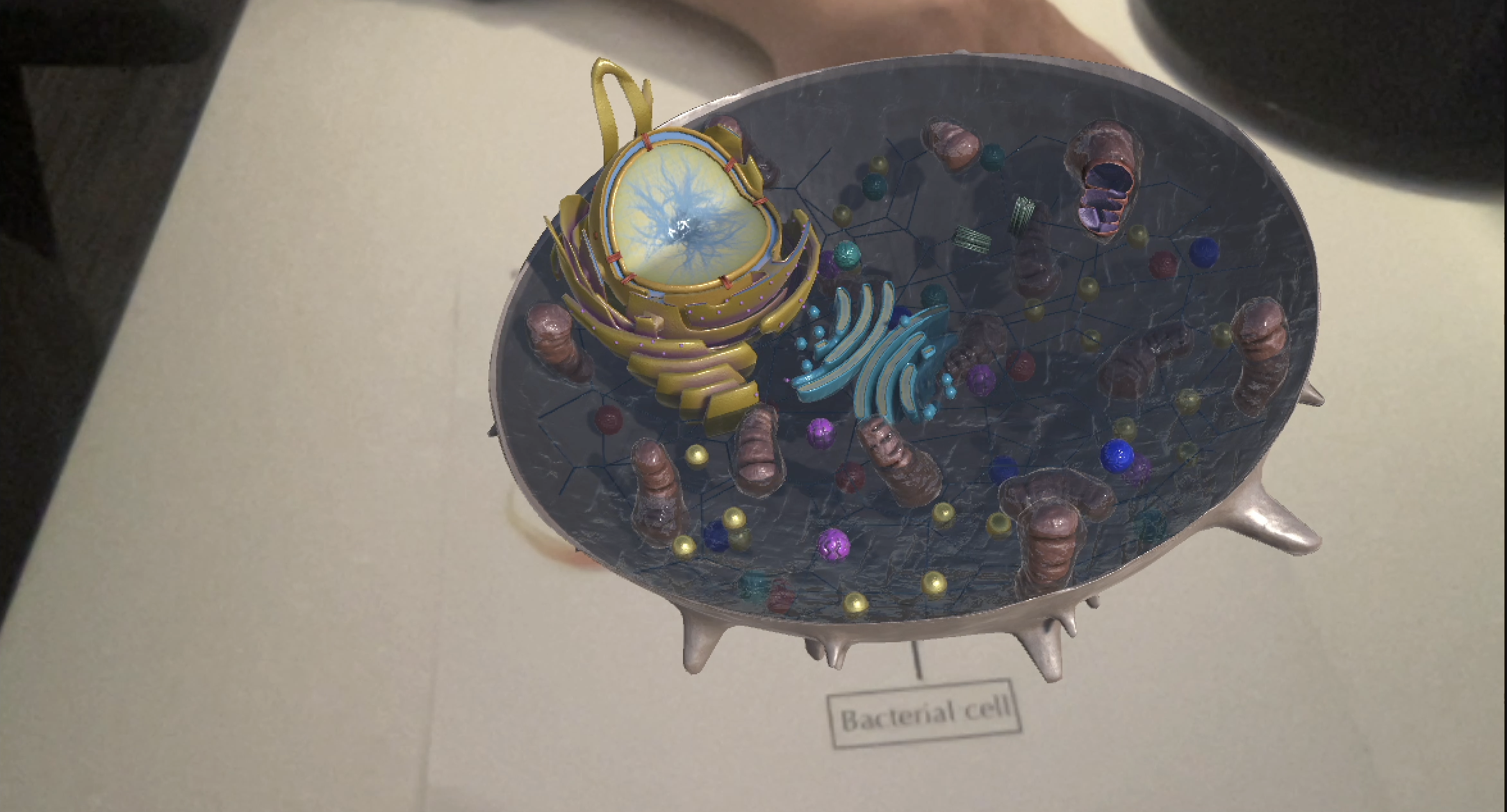
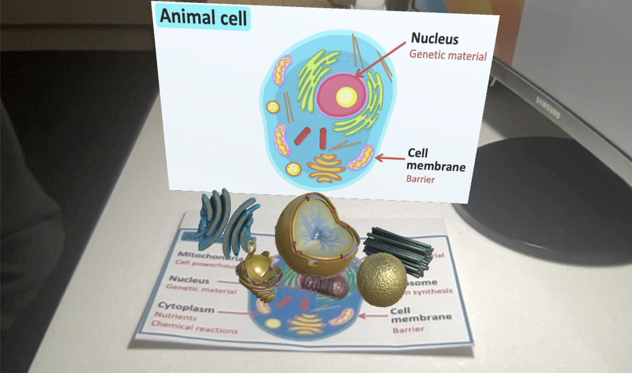
The augmented reality app displays four cell types: plant, animal, bacterial, and fungal cells. They display detailed cell compartments as well. Students can easily understand they reach cell has different cell compartments. To study more, plant cells contain organelles such as chloroplast, central vacuole, nucleus, reticulum, mitochondrion, and Golgi apparatus, and are represented as 3D models. However, animal cells also contain nucleus, mitochondrion, lysosomes, centriole, reticulum, and Golgi bodies. They have four common cell compartments which also function the same but indicate that plant cell has two different compartments that animal cell does not have by 3D models. It can be assumed that the users can actively engage in investigating more about cells by visual perception with 3D models.
6. Reference
I have used images and videos via the links below:
https://sciencenotes.org/plant-cell-diagram-organelles-and-characteristics/
For assets, I used “Biology Cells Pack” from System-IntegraTech.
Leave a comment
Log in with itch.io to leave a comment.